A quantitative electroencephalogram, or QEEG is an assessment tool that can be used to create brain maps. The QEEG measures the electrical activity of the brain. The resulting brain waves represent the brain’s internal communication system. Electroencephalography is typically used by neurologists to evaluate and diagnose seizure activity and epilepsy. Several other disorders such as narcolepsy and brain tumors are also assessed with an EEG.
The quantitative function of the QEEG is essentially the comparison of an individual brain’s electrical activity and brain wave expression against a normative database of thousands of other brains of the same age. The normative database includes brains that have no history of mental illness, seizure activity, addiction or head injury. This analysis reveals patterns of communication in the brain where the brain may be over or under functioning and these are often shown in brain maps. A QEEG-D is a specialist who is recognized to be qualified to review and interpret brain maps and brain wave activity.
The findings from the QEEG, as shown in brain maps can be used to better understand how brain functioning may be reflecting or contributing to symptoms associated with mental health conditions such as anxiety, depression, PTSD, traumatic brain injury, migraine, and ADHD among many others. Furthermore, these findings can be used to create neurofeedback and neuromodulation treatment protocols. By training the brain wave activity, through neurofeedback and neuromodulation, more optimal states of functioning within the brain can be established over time. These changes tend to be stable once well-established so further follow-up and training is rarely required. These changes are associated with a reduction in symptoms, improved functioning, and enhanced quality of life for patients.
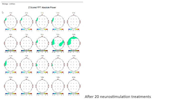
The following brain maps represent brain waves at each frequency of 1 hertz bins. This brain map is for someone with a history of concussion and the brain is functioning well below normal and reflects a brain that is shut down which is commonly seen with brain injury.

After a series of training sessions, the changes in the brain can be shown with brain maps and these changes are often associated with symptom reduction or symptom elimination. These images are the same brain after 20 sessions of neurostimulation. The patient reported greater energy and less anxiety and mood dysregulation.
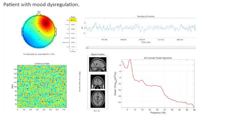
Other tools often used to assess brain functioning include Independent Component Analysis (ICA) and Event Related Potentials (ERP). Collectively, these tools provide a wealth of information about how the brain is functioning and what neuromodulation training can support improvements in sleep, mental clarity, energy, and mood regulation.


.jpg)